Abstract
Introduction Iron deficiency anemia (IDA) in autoimmune gastritis (AIG) is common. However, the development of IDA post-treatment of AIG with vitamin B12 supplementation remains underappreciated. Despite overlapping pathogenetic mechanisms directly linking AIG and the development of IDA, covert iron deficiency often lurks until repletion treatment. These patients thus experience late diagnoses, leading to a persistence of anemia symptoms and poor quality of life. Aim This case report details the development of IDA post-vitamin B12 supplementation in an AIG patient, highlighting the need for iron status evaluation in such patients. Case Summary A 42y.o female without known medical comorbidities was diagnosed with distal symmetric chronic sensory neuropathy due to vitamin B12 deficiency in PA. This was after she presented with longstanding lower limb numbness, paresthesias, headaches, palpitations, and light-headedness without weakness, arthralgia, constitutional symptoms or stigmata for connective tissue disease. She had no heavy menstrual bleeding, gastrointestinal concerns or substance use. Her diagnosis was confirmed following low serum vitamin B12 levels (75ng/ml), positive intrinsic factor antibodies (IFA) and macrocytosis on hemogram. She was initiated on replacement therapy, had improvement, and thereafter interval worsening of symptoms. Subsequent reassessment revealed microcytosis on hemogram, which prompted evaluation of iron studies, revealing concurrent IDA. She was treated with IV iron and continued her vitamin B12 replacement, with complete resolution of anemia symptoms 6 months later. Discussion This was a case of IDA that was unmasked by vitamin B12 replacement in a patient with AIG. The coexistence of IDA-AIG is common due to shared pathogenetic mechanisms. Noteworthy, 46% of AIG patients end up with a diagnosis of IDA at or within 1 year of diagnosis of AIG, highlighting the interaction of the two conditions. Conclusion IDA may present at diagnosis or during the treatment course for AIG. It is important to thus assess iron status in all patients with AIG for early diagnosis and intervention.
Keywords
Pernicious anemia Autoimmune gastritis Iron deficiency anemia unmasked.
Background
Iron deficiency anemia (IDA) accounts for up to 50% of anemias globally (1), primarily resulting from blood loss, with less contribution from poor dietary intake, absorptive pathologies, and increased demand states. It leads to poor quality of life and increased healthcare expenditure, necessitating prompt diagnosis and treatment (2). Pernicious anemia (PA) is a vitamin B12 deficiency caused by autoimmune destruction of gastric parietal cells, leading to loss or dysfunction of intrinsic factor (3). While IDA coexists with and worsens outcomes in other anemias, less is known about its relationship with vitamin B12 deficiency in PA.
While Carmel et al. initially described a 20.7% prevalence of IDA in patients with PA (1), Lenti et al. later suggested that up to 20% of patients with unexplained or refractory IDA have pernicious anemia (4), pointing to a possible bidirectional relationship. The mechanisms of IDA in PA involve achlorhydria and its effects on iron absorption, Helicobacter pylori colonization in atrophic gastritis, and the regulation of erythropoiesis (4, 5). Given the burden of IDA in PA and the likelihood that PA treatment may unmask underlying IDA, we highlight the need for regular iron status assessment in PA patients (6).
Our case report details the diagnosis and initial care of a patient with serologically confirmed PA, in whom initial vitamin B12 replacement therapy is suspected to have unmasked a concurrent IDA, leading to interval recurrence and worsening of anemic symptoms. She was subsequently successfully evaluated for IDA and treated concurrently with vitamin B12 supplementation, achieving symptomatic resolution, highlighting the role of iron status assessment in PA patients.
Case Summary
Patient Information: A 42y.o. female, para 2+0, without medical comorbidities, was initially evaluated for a 6-month history of progressive bilateral lower-limb numbness, with accompanying tingling and a burning sensation on the plantar surfaces. She reported no lower-limb weakness, swelling, knee or low back pain at the time, and had no history of joint pain, constitutional symptoms, skin rash/lesions, hair loss, menstrual changes, Raynaud’s phenomenon, digital ulcers, dry eyes, neck swelling, altered bowel patterns, or dyspepsia. However, she reported occasional headaches 2 months prior to presentation, with lightheadedness, palpitations, and lethargy, but no syncope, convulsions, effort intolerance, cough, or leg swelling. There was no history of abnormal menstrual bleeding, gastrointestinal surgeries, prior liver disease, prolonged proton pump inhibitor use, inflammatory bowel disease, or worm infestations. She worked as a client relations officer and had no history of alcohol use, substance abuse, or chronic prescription medications.
Clinical Findings: Examination revealed a fair-appearing, well-dehydrated, afebrile patient without pallor or scleral icterus. Vital signs: blood pressure 130/79 mmHg, pulse 79 beats/min, oxygen saturation 97% on room air, temperature 36.90C, and respiratory rate 14 breaths/min. Her abdomen was nontender, of normal fullness, moving with respiration, with clear hernial orifices. Neurologically, her Glasgow Coma Scale (GCS) was 15/15, with grossly intact cognition and intellect. Pupils were bilaterally equal and reactive. No obvious cranial nerve palsies were noted, and power, bulk, and tone were normal in both limbs. Clonus and Babinski were negative bilaterally, but fine touch assessment was equivocal. Bilateral +1 ankle reflexes were noted, with preserved knee reflexes. Coordination and proprioception were normal, as were cardiovascular and respiratory system assessments.
Diagnostic Assessment: Per Table 1 below, she had macrocytic anemia, low vitamin B12 levels, negative antiparietal cell antibodies (APCA), positive intrinsic factor antibodies (IFA), and elevated serum homocysteine at 23.8 umol/ml (5-15). Her erythrocyte sedimentation rate (ESR) was normal at 7 mm/hr (0-20), as were thyroid-stimulating hormone (TSH) 1.57 mIU/l (0.3-3.94), calcium 2.14 mmol/l (2.10-2.60), magnesium 0.81 mmol/l (0.7-0.9), and folate 33.95 ng/ml (7.2-34.8). She had a negative Helicobacter pylori antigen test, low vitamin D3 28.64 ng/ml (30-50), and a negative rapid plasma reagin (RPR). Her urea, electrolytes, and creatinine (UECs), urinalysis, liver function tests (LFTs), hemoglobin A1C (5.4%), antinuclear antibodies (ANA), lipid panel, basic toxicology, and virology panel were normal. Nerve conduction studies (NCS) were not performed due to cost and availability. An assessment of distal symmetric chronic sensory neuropathy with macrocytic anemia due to vitamin B12 deficiency was made.
| Category | Test | Result | Range | Status |
|---|---|---|---|---|
| CBC | Hemoglobin | 11.7 | 12–16.5 g/dL | Low |
| Mean Cell Volume (MCV) | 103 | 83–101 fL | High | |
| Platelet Count | 226 | 150–410 ×10³/µL | Normal | |
| White Blood Cell Count | 3.19 | 4–10 ×10⁹/L | Low | |
| Metabolic | Vitamin B12 | 75 | 150–800pg/ml | Low |
| Folate | 33.95 | 7.2–34.8ng/ml | Normal–High | |
| Homocysteine | 23.8 | 5–15 µmol/L | High | |
| Vitamin D₃ | 28.64 | 30–50 ng/mL | Low | |
| AIG panel | APCA | Negative | Negative | Normal |
| IFA (IgG) | Positive | Negative | Abnormal | |
| Inflammatory | ESR | 7 mm/hr | 0–20 mm/hr | Normal |
| Infectious screen | H.pylori Stool Antigen | Negative | Negative | Normal |
| RPR | Negative | Negative | Normal | |
| Thyroid Function | TSH | 1.57 | 0.3–3.94 mIU/L | Normal |
| UECs | Urea | 2.4 | 2–8 mmol/L | Normal |
| Creatinine | 77 | 65–120 µmol/L | Normal | |
| Sodium | 136 | 135–145 mmol/L | Normal | |
| Potassium | 4.1 | 3.5–5.5 mmol/L | Normal | |
| Chloride | 102 | 97–110 mmol/L | Normal | |
| Urinalysis | Specific Gravity | 1.020 | 1.005–1.030 | Normal |
| Protein | Nil | Negative | Normal | |
| Blood | Nil | Negative | Normal | |
| Leukocytes | Nil | Negative | Normal | |
| Glucose | Nil | Negative | Normal | |
| Bilirubin | Nil | Negative | Normal | |
| LFTs | AST | 16 | 8–40 U/L | Normal |
| ALT | 21 | 5–40 U/L | Normal | |
| Albumin | 37.9 | 35–45 g/L | Normal | |
| GGT | 67.57 | 0–50 U/L | High | |
| Total Bilirubin | 14.4 | 1.7–21 µmol/L | Normal | |
| Glycemic | Hemoglobin A1C | 5.4% | <5.7% | Normal |
| Autoimmune | ANA | Negative | Negative | Normal |
| Lipid Panel | Triglycerides | 1.02 | 0.4–1.81 mmol/L | Normal |
| LDL Cholesterol | 1.96 | 0–3.1 mmol/L | Normal | |
| HDL Cholesterol | 0.33 | 1.07–1.89 mmol/L | Low | |
| Other Panels | Toxicology Panel | Normal | — | Normal |
| Virology Panel | Normal | — | Normal |
Therapeutic Intervention: She was apprised of the chronic nature of her condition and the need for lifelong replacement therapy, and was initiated on a daily vitamin B12 nasal spray. She was also given a 3-month prescription for pregabalin 75 mg nightly and vitamin D supplementation (600 IU/day). At her 3-month review, her neuropathy had substantially resolved, and she could now sleep better and concentrate on her office tasks. Her repeat vitamin B12 level was 222 ng/mL and continued to rise, reaching a 12-month peak of 707 ng/mL (Figure 1). However, she subsequently stopped using the spray 3 months before her scheduled 18-month review, owing to concerns about ‘nasal discomfort’ felt each time she used it, and to new-onset malaise, palpitations, and difficulty concentrating at work, which she described as odd and not in tandem with her previous symptoms. The neuropathy, however, did not recur.
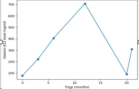
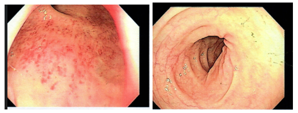
Further care and follow-up: At her 18-month review, her vitamin D3 level was 42.16 ng/mL, and the repeat vitamin B12 level was 90 ng/mL. Other electrolytes and the metabolic panel were normal. Her CBC, however, was now microcytic, with hemoglobin 11.5 g/dL (12-16.5), red cell volume 73 fL (83-101), platelet count 251 × 10^3/µL (150-410), and white cell count 6.19 × 10^6/µL (4-10). A concomitant iron deficiency was suspected to have been unmasked by the initial B12 treatment, accompanied by a now-relapsing B12 deficiency. Thus, confirmatory iron studies revealed a serum iron level of 4.51 µmol/L (5.83-34.5), transferrin 2.99 g/L (2.50-3.80), transferrin saturation 6.0% (20-50%), and ferritin 6.31 ng/mL (13-150). An esophagogastroduodenoscopy (EGD) follow-up after positive occult stool testing showed mild gastritis (Figure 2), and biopsy findings showed chronic pylori-negative gastritis without glandular atrophy, dysplasia, or intestinal metaplasia. Colonoscopy was normal.
She was initiated on IV ferric carboxymaltose on alternate days for 3 doses. Repeat ferritin level 1 month later was 134 ng/mL, with complete resolution of her fatigue. Additionally, Stherapy for vitamin B12 deficiency was commenced almost simultaneously, initially with intramuscular hydroxocobalamin 1000 mcg every other day for 2 weeks, then quarterly for life. Subsequent reviews have shown no concerns. B12 level 1 month post re-initiation was 309 ng/mL, with hemoglobin 12.9 g/dL (12-16.5), red cell volume 82.4 fL (83-101), platelet count 156 × 10^3/µL (150-410), and white cell count 4.19 × 10^6/µL (4-10).
Discussion
Vitamin B12 deficiency and PA are hallmarks of autoimmune gastritis (AIG). In this case, the initial diagnosis of PA was based on anemic symptoms (lethargy, palpitations, and effort intolerance), low B12 levels, elevated serum homocysteine, and a positive IFA, and treatment was appropriately initiated. However, initial improvement followed by interval worsening of the patient's status and the development of microcytosis necessitated re-evaluation of the coexistence of IDA and PA. Notably, while B12 deficiency has long been established as the major consequence of AIG, achlorhydria and intrinsic factor loss or dysfunction also cause iron and other micronutrient malabsorption (6). Consequently, iron and other micronutrient deficiencies may ensue, necessitating a broader assessment and care.
Characteristically, IDA may be diagnosed before or during the initial PA diagnosis in about 20%-52% of cases, or within the first fourteen months of PA diagnosis in 23%-46.4% of patients (1, 3). Rogez et al., in their PA cohort study, reported iron deficiency in 46.4% of newly diagnosed PA patients and in 28.3% during follow-up (6). This emergence of concurrent or unmasked IDA in PA patients is hypothesized to be linked to AIG disease duration, severity, and molecular dynamics that influence disease evolution (7).
An initial concurrent diagnosis often points to longstanding PA with established malabsorptive effects, and microcytosis from IDA or normocytosis can mask and delay the diagnosis of B12 deficiency (5). Iron deficiency in this segment results from reduced iron absorption due to longstanding achlorhydria from gastric parietal gland atrophy (5). Thus, hypothetically, vitamin B12 replacement before correction of IDA could further worsen iron deficiency (7).
In later diagnosis, also called unmasked iron deficiency, as in our case, IDA development is thought to be a consequence of improved erythropoiesis after vitamin B12 supplementation (2). Here, renewed erythropoiesis consumes the already meager iron stores, causing a further decline in iron levels and leading to symptomatic IDA.
IDA treatment involves iron supplementation. The choice between oral and intravenous iron formulations depends on availability, tolerability, cost, and comorbidities (8). In our case, the oral formulation was avoided because of dyspeptic concerns, impaired absorption, and the need to rapidly correct the anemia. IV iron has been shown to increase hemoglobin, serum iron, and ferritin levels more than oral iron, but it does not reduce the need for transfusion (8, 9).
Conclusion
In summary, our case report affirms that IDA should be suspected and actively evaluated in all patients with PA. The assessment should include routine iron tests (serum iron, ferritin, and transferrin saturation), and alternative causes of IDA should be excluded. Notably, treatment should be initiated concurrently with B12 replacement therapy for symptomatic resolution.
Source of Funding
No funding was used for this research.
Availability of Data
The patient data used in advancing the case review can be requested from the authors.
Conflict of Interest
The authors declare no conflict of interest.
References
- Carmel R, Weiner JM, Johnson CS (1987). Iron deficiency occurs frequently in patients with pernicious anemia. Jama. 257(8):1081-3. Available from DOI ↗ Google Scholar ↗
- Longo DL, Camaschella C (2015). Iron-deficiency anemia. N Engl J Med. 372(19):1832-43. Available from DOI ↗ Google Scholar ↗
- Cimadevila GC, Thain A, Dib MJ, Ahmadi KR (2024). Investigating The Causal Link Between Serum Iron Status and Pernicious Anaemia Risk: A Mendelian Randomisation Study. medRxiv. 2024-10. Available from DOI ↗ Google Scholar ↗
- Lenti MV, Rugge M, Lahner E, Miceli E, Toh BH, Genta RM et al. (2020). Autoimmune gastritis. Nature Reviews Disease Primers. 6(1):56. Available from DOI ↗ Google Scholar ↗
- Remacha AF, Sarda MP, Canals C, Queralto JM, Zapico E, Remacha J et al. (2013), Combined cobalamin and iron deficiency anemia: a diagnostic approach using a model based on age and homocysteine assessment. Annals of hematology. 92(4):527-31. Available from DOI ↗ Google Scholar ↗
- Rogez J, Urbanski G, Vinatier E, Lavigne C, Emmanuel L, Dupin I et al. (2024). Iron deficiency in pernicious anemia: Specific features of iron deficient patients and preliminary data on response to iron supplementation. Clinical Nutrition. 43(4):1025-32. Available from DOI ↗ Google Scholar ↗
- Zulfiqar AA, Pennaforte JL, Drame M, Andres E (2013). Pernicious anemia and iron deficiency: Possible link?. European Journal of Internal Medicine. 24:e162-3. Available from DOI ↗ Google Scholar ↗
- Bharti V, Katiyar DK (2025). Comparative Study of Oral vs. Intravenous Iron Supplements in the Management of Anemia in Chronic Kidney Disease. J Neonatal Surg [Internet]. 14(12S):562-7. Available from DOI ↗ Google Scholar ↗
- Das SN, Devi A, Mohanta BB, Choudhury A, Swain A, Thatoi P (2020). Oral versus intravenous iron therapy in iron deficiency anemia: an observational study. Journal of family medicine and primary care. 9(7):3619-22. Available from DOI ↗ Google Scholar ↗